Thursday 19 October 2023

Self-indulgent re-branding exercise

 Introducing: Enjoying Field Mycology ...


Hello, reader! It is always a pleasant surprise to receive any indication that real human beings with whom I might have something in common are reading this blog, as I did recently. 

Somebody was kind enough to comment that perhaps 'misidentifying fungi' is a misnomer for the kind of content I've been sharing with the world the past few years. So ... time for a rebrand!

You can find my latest post over on substack: 

https://enjoyingfieldmycology.substack.com/

(I don't really know what I'm doing or where I'm going with this, so bear with...)


Ta ta for now, and thanks for reading my blog.

Clare

Saturday 8 April 2023

Rusty Threefer


'Tis the season for having a go at botany: one fungal substrate at a time. 

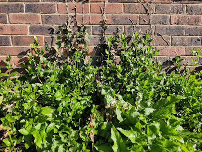
If you zoom right in on this sickly-looking Periwinkle (Vinca sp.) then you can just make out a hairy edge to the leaves...

I think this makes today's plant Greater Periwinkle Vinca major

And if I've got that right, then I think those black pustules scattered generously across its leaves are one of the life stages of the rust fungus Puccinia vincae

Under the stereo microscope (image below) you can see the hairy margin of the leaf nicely and I think the orange specks are 'stage 0' of the fungus's lifecycle: the spermogonia. The black specks are 'stage I': the aecia.

One or two of the leaves are showing these gingery-coloured pustules, 'stage II': the uredinia.

That gingery-coloured stuff is actually a mass of urediniospores, pictured here at 100x magnification (image is of a cross-section through one of those pustules):

This is what the urediniospores look like at 40x magnification.

I really had to hunt for 'stage III', but eventually managed to track down a few solitary teliospores (one pictured below at 1000x magnification).


I read on the fungi.org.uk forum that the teliospores of Puccinia vincae look like peanuts, which gave me some motivation to fiddle around with oil immersion and a bit of focus-stacking. And I have to agree, it does look like a peanut.

***

That might have been the end of my study of Puccinia vincae but, what do we have here? 


Some ashy-grey looking pustules (or, 'sporodochia'?):

I think this gives us the threefer! (If I am correct) what we have here is another fungus: Tuberculina sbrozzii, parasitising the rust fungus Puccinia vincae which is itself a biotrophic parasite on the Greater Periwinkle Vinca major 

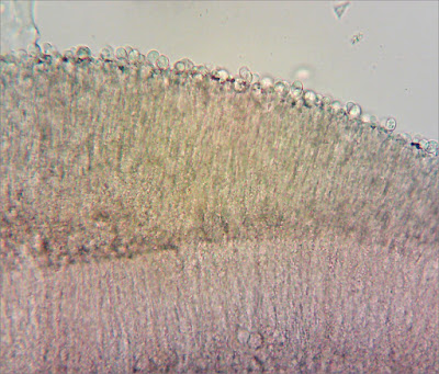
These ashy-grey sporodochia are structurally very different to the uredinia I was looking at earlier. Here is an image of one in cross-section at 100x magnification. 

The outer surface is covered in little round conidia.

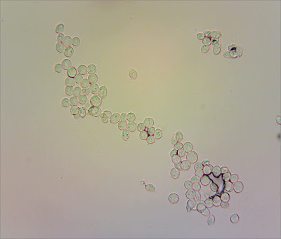
Here are a load I scooped out from the middle of a sporodochium, at 400x magnification:

You can see that they are clear, or 'hyaline', and when I measured them they were about 10 microns in diameter - which is the right ballpark for Tuberculina sbrozzii.


For the record

Date: 8 April 2023

Private site in East Sussex

(Records will be entered into iRecord in due course.)

Sunday 19 March 2023

Spring is in the air (and in that pile of decaying debris)

At Sussex Wildlife Trust's Woods Mill nature reserve, at the far end of the lake, near the stream, there is a pile of decaying flood debris. Little bits of sticks and leaves, and old bits of reeds and sedge. On top of this pile there was one thing that was covered in miniscule white specks:


 ... a decaying catkin. After close inspection, I'm pretty sure it's one of last year's Goat Willow catkins. 

Photo of Goat Willow catkin, borrowed from tcv.org.uk: https://treegrowing.tcv.org.uk/identify/goatwillow

The white specks swarming over the dead catkin didn't look like much to the naked eye. They didn't even look like much under a hand lens. But under the stereomicroscope they are rather lovely.

The fertile surface of the tiny cup-shaped fruit bodies (none of them more than 0.5mm across), fringed with hairs, fairly sparkle under the light. 

The whole outer surface is covered in hairs, giving a downy appearance.

Under the microscope, they've got avocado shaped spores.


... and the asci, stained with Lugol's iodine, are showing a blue (amyloid) reaction at the tip.


It's been a long time since I looked at ascos, but I think that means the asci are inoperculate and the bit that's turned blue is the apical ring. 

My tech set-up isn't fully cooperating this evening, so I haven't been able to measure anything...  

But I reckon I might be able to identify this collection??? Cyathicula amenti (= Crocicreas amenti) fits the habitat and morphological characteristics. And according to 'Microfungi on Land Plants' by Ellis & Ellis, it's "common" in March to April.


For the record

Date collected: 18 March 2023

Location: Woods Mill, West Sussex


Sunday 23 October 2022

To Ditchling

After a chaotic week, I failed to check either the weather forecast or my emails ahead of the Sussex Fungus Group survey at Ditchling Beacon this morning; so I didn't know torrential rain & thunderstorms were forecast and it was cancelled. 

Hence I was at the bottom of Ditchling Beacon this morning ... and the rain seemed to have slackened off for a bit ... I thought  I'd go for a mooch around the old chalk pits.

Most of the mushrooms I saw looked completely mullered, but a plucky little white waxcap had weathered the previous night's storms. 


No scent, so it was one of those Snowy Waxcap Cuphophyllus virgineus ones.

I almost missed this moss-coloured mushroom, nestled amongst some longer vegetation...

Those are the distinctive tones of Mouse-pee Pinkgill Entoloma incanum. The yellow-green stipe and bluish-green staining on this species when bruised (which you can just about see in the second photo below, where the stipe meets the gills) is just WILD. I love it. Makes this mushroom look like a total raver.

But I found out earlier this week ID'ing E. incanum isn't quite so simple any more...

Given my penchant for grassland fungi, I couldn't resist placing an order for the new Entoloma book by Machiel E. Noordeloos and it arrived on Tuesday from Summerfield Books. It's a beast!

On page 259 (of 968 !!!), Noordeloos explains: 

"Entoloma incanum is one of the iconic species of subgenus Cyanula with its yellow-green tinges, blue-green discolouration at the stipe base and strong smell, which reminds of that of mouse-nests. Until recently the species was therefore easily identifiable in the field. However, during the phylogenetic study of the subgenus Cyanula, two genotypes fitting the current morphological concept of Entoloma incanum were revealed ... indicating that we have two different species in Europe. As a result, E. verae was published (Crous et al., 2021) as new to science."

He goes on to explain that the two species are thought to be separable on spore size. 

Happily, Noordeloos's book also includes guidance on how to measure Entoloma spores, because this has always been a mystery to me. 

Figure from Fungi Europaei: Entoloma s.l. 5B by M.E. Noordeloos (2022). (c) Lidia Carla Candusso.

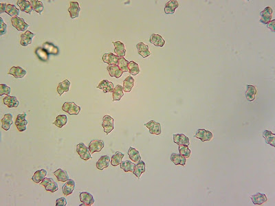
So this is what the spores from my collection look like:

And these are my measurements:

So I think this makes my collection Entoloma incanum sensu stricto.

***

On one of the banks in the old chalk pits, I found the remains of a mushroom I thought might have been one of those nice 'big blue pinkgills'. But not enough of it to make me want to attempt an ID!

Then Martyn Ainsworth appeared ... so I seized my opportunity to cross-question him about the  Cuphophyllus I had found earlier (first two photos, above). 

I wondered if I should be calling it "var. fuscescens'... But Martyn explained that phylogenetic work (in progress) has shown that "Cuphophyllus virgineus sensu lato" clusters into multiple clades and collections with a brownish central spot on the cap show up in several of these clades. So I can call it var. fuscescens (as described in Boertmann's The Genus Hygrocybe, 2nd Edition) if I want to – but it sounds like Science is going to tell us that these visual characters / morphological concepts don't map across neatly to DNA-based species concepts (so those little brown spots in the centre of the cap might turn out to be kind of irrelevant for species-ID / taxonomic purposes). 

I think I'll keep things simple and call it Cuphophyllus virgineus s.l..

***

Martyn recalled that there's a field towards the bottom of Ditchling Beacon where he's found Entoloma bloxamii before, so we went to check that out. 

 We found loads! Reckon we must have seen twenty, at least.

Collection 1. Spores shown below.

With those slight lilac tones, Martyn advised that a check on spore size could confirm the ID as Entoloma bloxamii sensu stricto. (For more info see the paper from Field Mycology on Big Blue Pinkgills, here.)


In E. bloxamii, the spores often exceed 8 μm. That certainly looks to be the case for my collection 1, above.

I looked at a few different collections, pictured below in terrible light, and they all seemed to be showing similar colouration: greyish and slightly lilac tones (not the dark or strikingly blue tones of E. atromadidum / madidum).  

So I think what we found was a big population of E. bloxamii sensu stricto.

***

There were a few other species showing in this field. 

Toasted Waxcap Cuphophyllus colemannianus, with its distinctive intervenose gills. 


More Snowy Waxcap Cuphophyllus virgineus, with and (as pictured below) without brownish central spot.

 
Plus some kind of Entoloma sp. with mycenoid cap shape and heterodiametrical (?) spores.


I hoped the mycenoid (almost campanulate?) shape to the cap and the adnate gills with a decurrent margin might be distinctive enough to allow for an ID based on field characters. But I think I'll have to summon up the courage to try and run this through Noordeloos's key, if I'm going to get anywhere with it.

***

One advantage of the terrible weather forecast ... had almost the whole place to ourselves. This never happens!


For the record

Date: 23 October 2022

Location: Ditchling Beacon

Records will be submitted via iRecord in due course. 

Saturday 17 September 2022

Fingal's Mushroom

Content warning: I make a lot of wrong assumptions about the identity of the mushroom in this blog, before I eventually come to a conclusion I have a reasonable amount of confidence in.


The main reason we were on the Isle of Iona (see previous post  about its waxcaps) was to take a trip to Staffa.

View of Staffa as the boat approaches from Iona.

The boat trip from Iona gives you one hour to explore the island which rises out of the North Atlantic Ocean like a giant basalt cathedral. One hour.

While our shipmates formed an orderly queue to see Fingal's Cave, we raced up the steps to explore the island. Surrounded by a dramatic, rocky coastline, Staffa has a grassy, heathy cap which I figured MUST have some mushrooms on it. 


At the side of an inauspicous-looking path, I spotted one small patch of waxcaps...

... hiding in the long grass.

Bright sunshine illuminated the scales on the cap of this little red mushroom. 

Underneath, pale yellow (adnate / adnexed?) gills, which looked almost white in the sunshine, contrasted with the red cap and smooth stipe. 

There was no time for closer observation in the field. We had to leg it back down to sea level or we'd miss our chance to see Fingal's Cave and the boat back to Iona. 

Fingal's Cave was amazing and impossible to photograph in five minutes with a phone, and the light outside blazing...

Returning to the boat we noticed the reddish-brown jellyfish hanging in the water, close to where it laps against these basalt columns. But there was no time to stand and stare, or get a better snap.

Back on the boat, I remembered the mushrooms I'd hurriedly thrust into my pocket earlier on. 

One specimen looked to have dark grey scales at the centre of the cap. 

Being in my pocket hadn't done them any good, but I could still make out this specimen has adnate gills. 

Hoping no one was watching, I gave them a good sniff. Nothing. 

I thought they were probably Hygrocybe miniata and, reading back through the descriptions in Boertmann's book (The Genus Hygrocybe, 2nd Edition), they are described as having "squamules concolorous and becoming yellow after slight drying, very rarely dark greyish in the centre" - which sounded about right. That species has distinctively pear-shaped ('mitriform') spores so I thought I'd take a look to confirm.

I'm still struggling with preparing microscope slides with dried material - and these were particularly squashed and broken by the time I got them home - so I tried a new approach: putting a drop of ammonia on a slide and just washing a couple of gills in it, to see if some spores would float off. It worked!

The following images are all captured at 400x magnification.


I found plenty of spores floating around. But none of them look mitriform to me. So I think that rules out H. miniata.

Hmm. 

I started again from the beginning with Boertmann's key, and found myself getting into some seriously tricky territory - these little red waxcaps can be rather cryptic. 

Could I have found Hygrocybe calciphila? Spore shape appears to be a better match. Boertmann says it's "generally rare, but may be overlooked" and describes it as known to occur on "basic (basalt or gypsum) soils"; I think it's probably a safe bet that the soil on Staffa is basalt-y!

Hygrocybe calciphila seems like the best fit with everything I've observed. But given it's so rarely recorded (and a new species to me) it would be good to get it confirmed by someone who knows what they're doing. Or perhaps an ITS barcode... 

Update 24/09/2022

I got an email from Andy McLay, Natural England's waxcap expert, advising me to compare my collection with Garlic Waxcap Hygrocybe helobia.  I think I ruled this out because I didn't get the faint smell of garlic ... but that was really an oversight on my part. 

Hygrocybe helobia is fairly distinct in its microscopic characteristics, having a regular gill trama with long, slender elements. 

I should get in the habit of checking gill trama characteristics - looking at a cross-section of the gills in thin-section - but the truth is I'm not very good at preparing the thin-sections so I avoid doing it. 

I am reasonably confident, having spent quite-a-long-time looking at some squashes from my collection under the microscope, it has a regular gill trama. 

Gill trama, from dried material. 400x magnification mounted in ammonia and stained with Congo Red.

So I think there is good evidence here for going with Andy McLay's suggestion that what I've got here is Hygrocybe helobia. That would fit with the spore shape being rather variable. Another feature of H. helobia that Boertmann mentions in his description is that the flesh is very fragile - that's something I observed as my collection broke into pieces almost as soon as I touched it.

Thanks Andy! 

***

Couldn't resist one more try at getting a decent gill trama squash. I think this one's better:

100x magnification. Gill cross-section mounted in water and stained (carefully!) with Congo Red.

Same as above, at 400x magnification. I am confident I can see long slender elements here, with tapering ends.


For the record

Date: 5 September 2022

Location: Staffa

Record will be entered into iRecord in due course, if I get a decent fix on what species it is!